
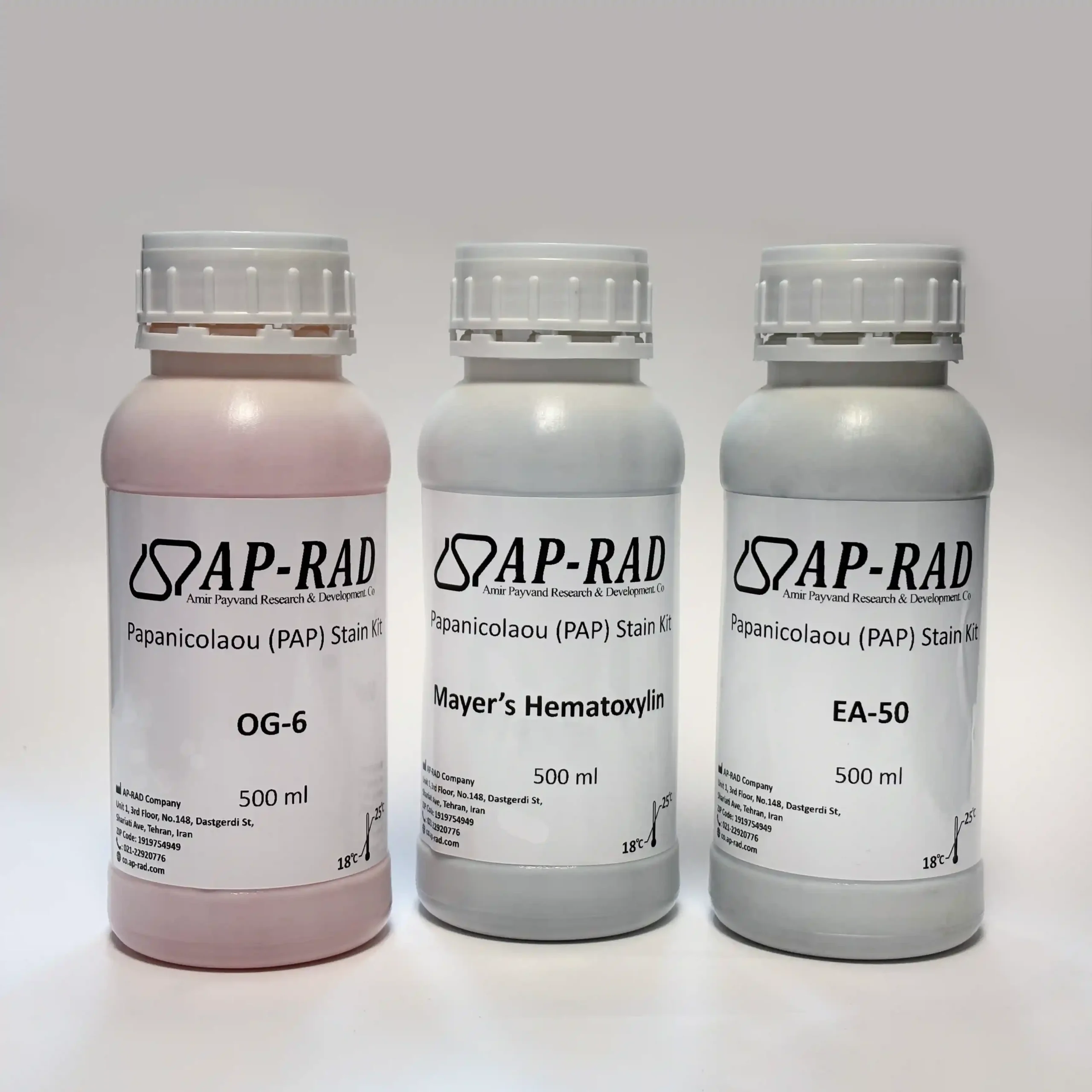
Papanicolaou (PAP) Stain Kit

Features:

Consists of 3 solutions, which produce 6 different colors in the sample:

  • Chromatin appears blue
  • Keratin appears orange
  • Superficial squamous cells, nuclei, cilia, and red blood cells appear in various shades of pink
  • The cytoplasm of metaplastic cells appears in various shades of blue-green

Description

Papanicolaou (PAP) Stain Kit | Papanicolaou Stain Kit

The Papanicolaou (PAP) Stain Kit clearly distinguishes between basophilic and acidophilic cellular components and displays a precise chromatin pattern. This kit is intended for use by laboratory professionals for staining smears.

The stained smear is examined to identify abnormal, inflammatory, and malignant cells (for example, in the diagnosis of cervical and uterine cancer). With this staining, chromatin appears blue, keratin appears orange, superficial squamous cells, nuclei, cilia, and red blood cells appear in various shades of pink, and the cytoplasm of metaplastic cells appears in various shades of blue-green. PAP staining consists of 3 solutions, which produce 6 different colors in the sample. Mayer’s Hematoxylin is a nuclear stain, while OG-6 and EA-50 are cytoplasmic stains.

The Papanicolaou (PAP) Stain Kit from ap-rad is designed and manufactured for use by laboratory professionals for staining smears.
Related Products
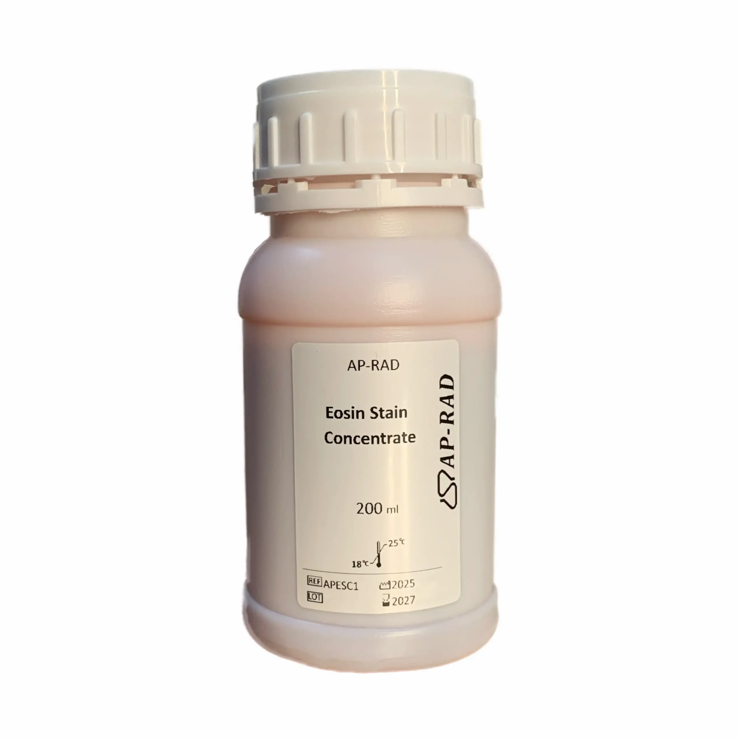
# Eosin Stain Kit
REF: APESC1
RUO
In the field of hematology, there are generally various staining kits and solutions that fall under the category of Romanowsky stains (blood staining with dyes containing thiazine and eosin).
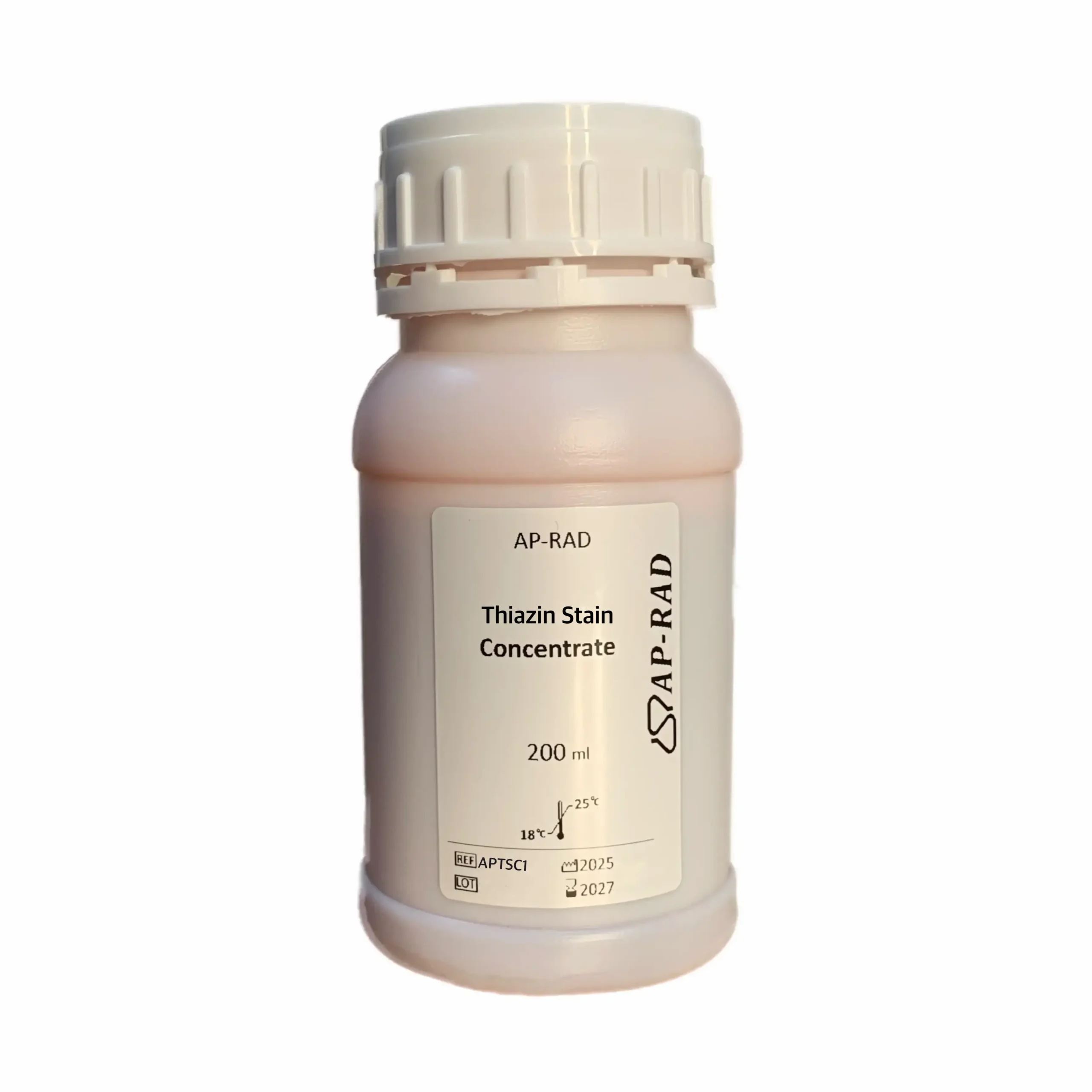
Thiazine Stain Concentrate Hematology Reagent
REF: APTSC1
RUO

Staining of:

  • Peripheral blood smears
  • Bone marrow aspirate smears
  • Body fluids
  • Sputum and other samples
# Myco-Prep Kit
REF: APMYPK
RUO
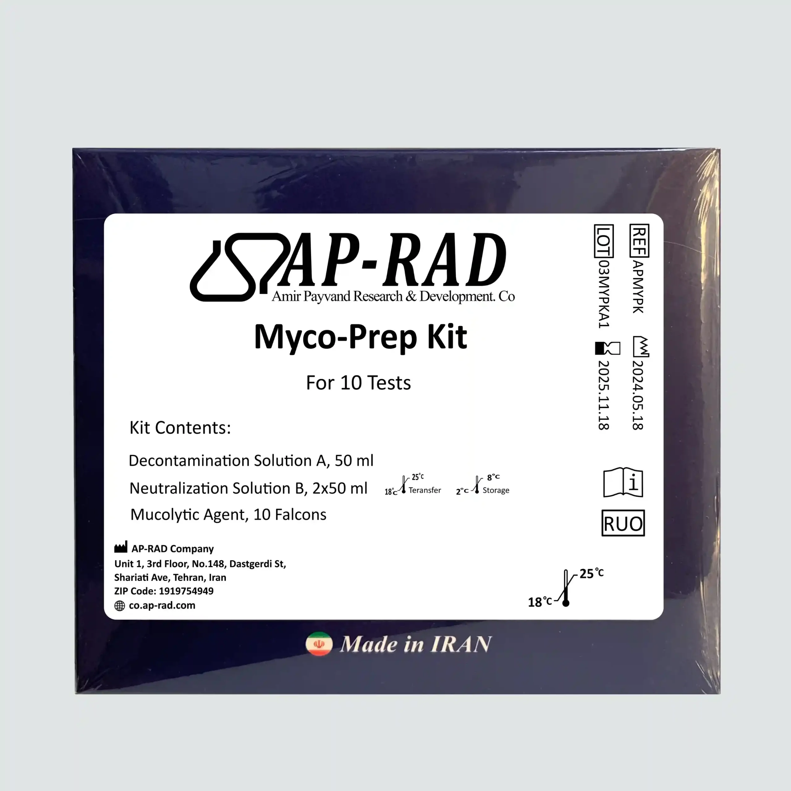
This kit is used for preparing sputum samples for the diagnosis of mycobacteria in Acid-Fast Bacilli (AFB) staining or TB Fluorochrome staining, culture, nucleic acid testing, and other related tests.
Bone Marrow Decalcifier Solution
REF: APBMDSK1
RUO
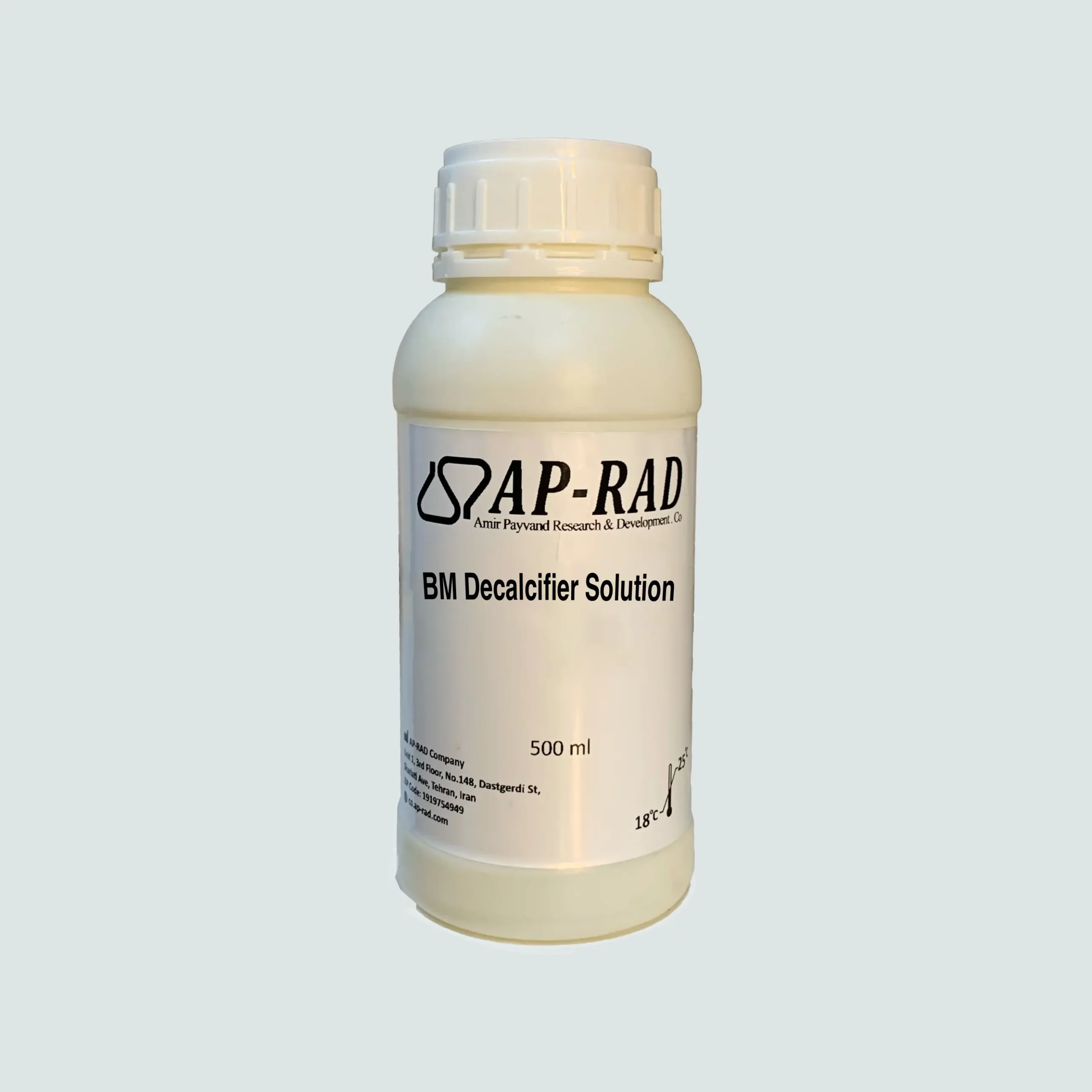
Removal of calcium from bone marrow in tissues to prevent interference in the testing process and subsequent analyses.
Periodic Acid Schiff (PAS) Stain Kit
REF: APPASSK1
IVD
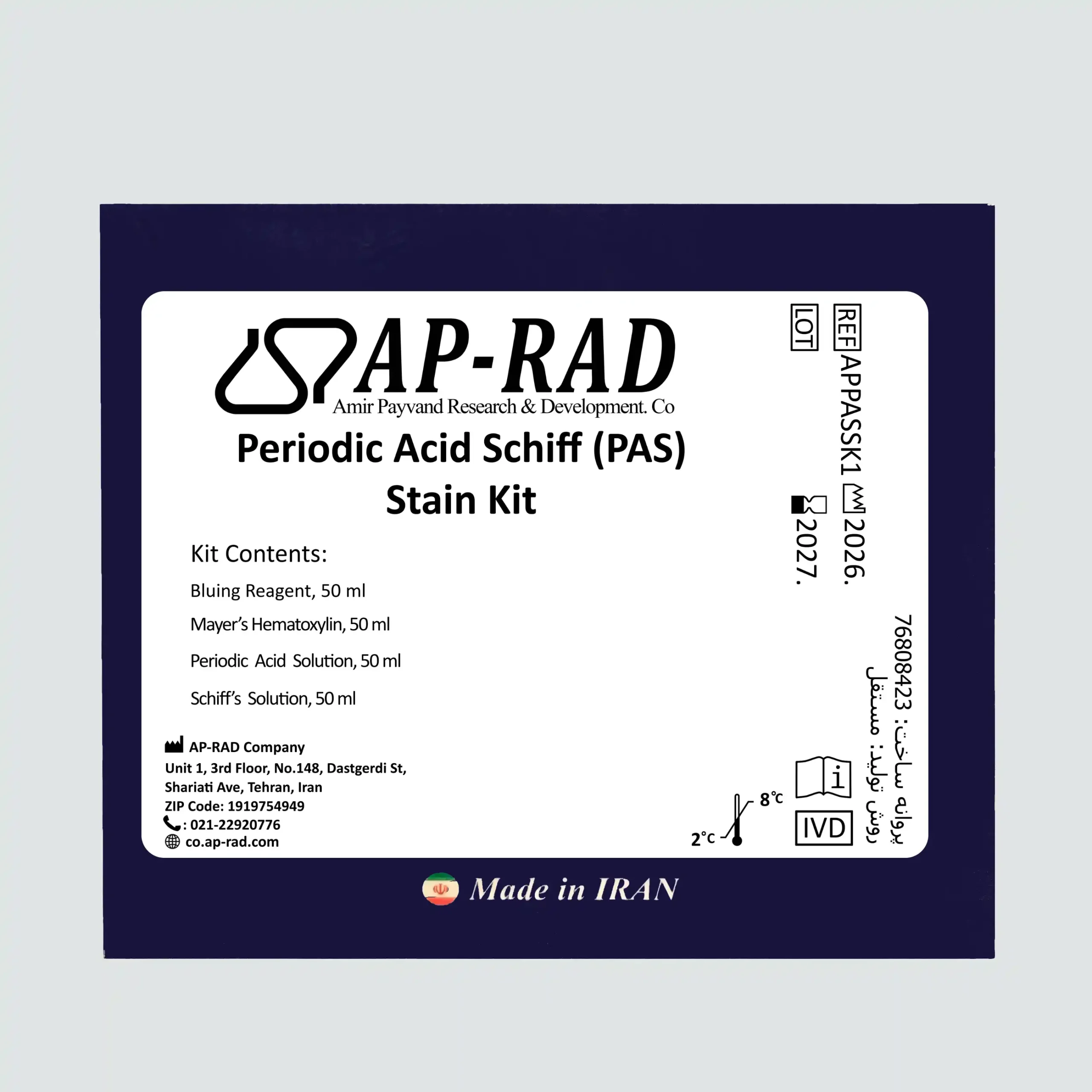
Periodic Acid Schiff (PAS) is a staining method used to identify polysaccharides such as glycogen and mucosubstances such as glycoproteins, glycolipids, and mucins in tissue. Additionally, this staining may be used to demonstrate fungal organisms in tissue samples.
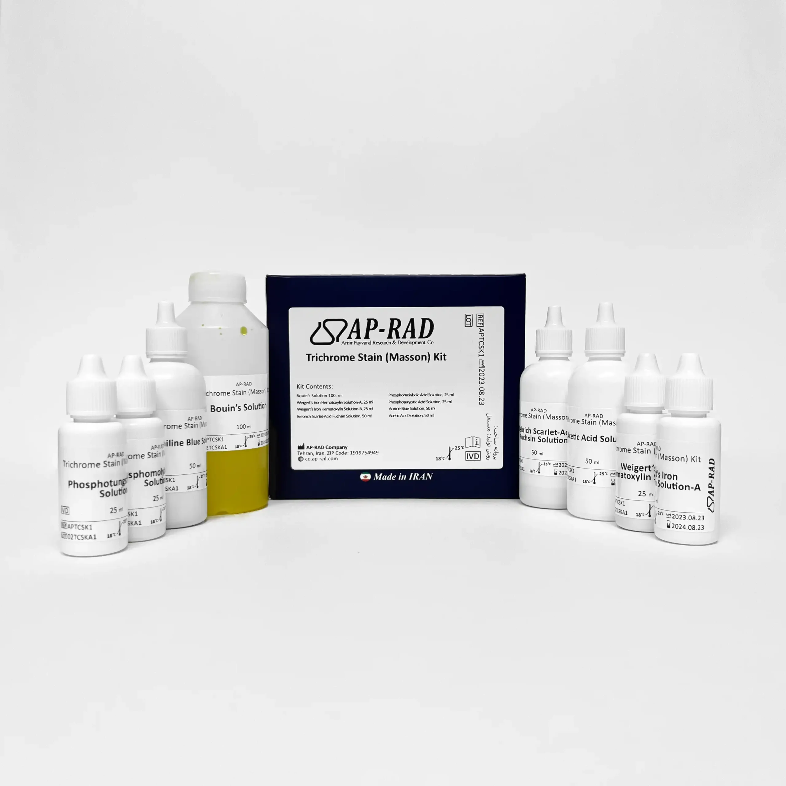
Masson's Trichrome
REF: APTCSK1
IVD
Masson’s Trichrome staining is a histological staining method used for the selective staining of collagen, collagen fibers, fibrin, muscles, and red blood cells.